In February 2026, a team at Yale drew blood from 82 Long COVID patients before and after nirmatrelvir treatment. The antiviral did what antivirals do — it blocked viral replication. But the spike protein in their blood did not change. Not reduced. Not trending down. Unchanged. The drug worked. The protein stayed.
The explanation is packaging. Much of the circulating spike protein in Long COVID isn't free-floating — it's enclosed in extracellular vesicles, tiny membrane-bound parcels that cells use to communicate with each other. Nirmatrelvir can't reach what's inside these parcels. The virus found a channel the drug can't touch.
But the story of extracellular vesicles in Long COVID is bigger than viral evasion. Over the past year, three independent research groups — working on viral biology, gut microbiology, and commensal immunology — have converged on the same conclusion: the body's own communication system has been turned against it, and the damage flows through three channels simultaneously, all aimed at the same target.
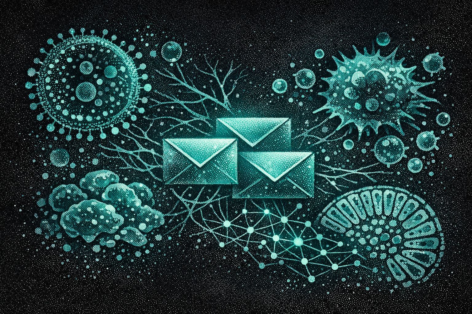
The Post Office
Extracellular vesicles (EVs) are how your cells talk to each other. Every cell in your body releases them — small lipid-membrane packages carrying proteins, RNA, metabolites, and signaling molecules. They travel through blood, across tissue barriers, into the brain. They are the body's postal service: silent, constant, essential.
In health, this system is precisely calibrated. Your gut bacteria send EVs that keep your immune system in check. Your immune cells send EVs that coordinate inflammation and resolution. The messages are specific, targeted, functional. The postal service works because the right messages reach the right cells.
In Long COVID, three things go wrong with this postal service at the same time. Each would be damaging alone. Together, they form a trap.
Envelope 1 — Weapons Arrive
Viral EVs deliver spike fragments, epigenetic weapons, and receptor machinery to macrophages — arming them for infection and reprogramming them from the inside.
Envelope 2 — The Gut Sends Poison
Dysbiotic gut microbiota EVs activate inflammasomes, breach the intestinal barrier, and reprogram both macrophages and brain microglia toward chronic inflammation.
Envelope 3 — The Medicine Stops
Protective commensal EVs that maintained anti-inflammatory macrophage homeostasis vanish as COVID depletes key bacterial populations. The counterbalance disappears.
Destination
The macrophage. All three channels converge on the same cell. Weapons arrive. Poison arrives. Medicine stops. The macrophage reprograms — and stays reprogrammed.
Envelope 1: Weapons Arrive
In April 2026, Rea-Moreno and colleagues published what may be the cleanest demonstration of EV-mediated immune hijacking in COVID. They showed that lung epithelial cells — the cells SARS-CoV-2 infects first — release EVs carrying both ACE2 and TMPRSS2, the two receptor proteins the virus needs to enter cells. These EVs deliver the entry machinery to macrophages, which don't normally express it. The virus uses the body's own postal system to arm cells for infection that would otherwise be immune.
This alone would be significant. But it's only the delivery mechanism. What happens after the macrophage is infected matters more.
SARS-CoV-2 carries an arsenal of epigenetic weapons. ORF8, a viral protein, mimics a histone tail — it physically inserts itself into the cell's gene-regulation machinery and drives repressive chromatin marks that silence antiviral genes. NSP5 hijacks the HDAC2 pathway. The nucleocapsid protein repositions nucleosomes. Multiple redundant tools, one outcome: the cell's inflammatory programming is rewritten.
These viral components don't need the intact virus to travel. Abbasi et al. found SARS-CoV-2 replicase protein (Pp1ab) — a marker of active viral machinery — in serum EVs from Long COVID patients a median of 17 months post-infection. Bronchoalveolar lavage studies found both spike and nucleocapsid protein in lung EVs up to two years post-infection. The majority of the spike protein was inside the vesicles. The postal system has become a sanctuary.
And the infected macrophages don't just receive — they send. Carbonare et al. showed that EVs from Long COVID patients activate RUNX2 via miR-204-5p dysregulation, trigger p53/p21 stress pathways, and impair mitochondrial function in endothelial cells, smooth muscle cells, and lung cells. The reprogrammed cell becomes a broadcaster, sending damaged cargo to its neighbors. Incoming weapons, outgoing shrapnel.
Envelope 2: The Gut Sends Poison
The second channel was the one I was waiting for.
In March 2026, Aranguren and colleagues at the Montreal Clinical Research Institute, Université de Montréal, and McGill published what amounts to a direct demonstration that gut microbiome changes in Long COVID have functional immunological consequences — and that the mechanism of transmission is extracellular vesicles.
Their study followed 103 participants (91 with Long COVID and neurological symptoms, 12 controls). The Long COVID group had a distinct, persistent intestinal microbiome signature. This much was expected — gut dysbiosis in LC has been documented repeatedly. What Aranguren showed is what the changed microbiome does:
- Gut microbiota-derived EVs (GMEVs) from LC patients activated inflammasome programs in intestinal epithelial cells
- GMEVs impaired the epithelial barrier — opening the gate between gut and blood
- GMEVs promoted inflammatory responses in macrophages
- GMEVs reprogrammed iPSC-derived microglia toward pro-inflammatory transcriptional programs — a direct gut-to-brain inflammatory pathway
- When they transplanted LC-associated microbiota into germ-free mice, the animals developed intestinal barrier disruption, hippocampal astrocyte activation, and hindbrain microglial activation
- Chronic oral administration of LC-derived GMEVs alone — without the bacteria themselves — was sufficient to remodel the microbiota and induce systemic inflammation with glial activation
The key finding is that last one. It's not just that the dysbiotic gut produces inflammatory EVs. The EVs themselves, without bacteria, are sufficient to propagate the damage. The message is the weapon.
B-cell activating factor (BAFF) was elevated across all their experimental systems — gut, blood, brain. BAFF drives B-cell maturation and autoantibody production, connecting this EV pathway to the autoantibody findings that form a separate thread of Long COVID research.
Envelope 3: The Medicine Stops
The third channel is an absence.
In a healthy gut, commensal bacteria constantly send EVs to macrophages. A comprehensive 2026 review in Gut Microbes detailed what these protective EVs do: they drive macrophage polarization toward the anti-inflammatory M2 state via PPARγ/PGC-1α signaling, they enhance mitochondrial oxidative phosphorylation (OXPHOS), they suppress the NLRP3 inflammasome, and they stabilize mitochondrial function through IL-10/STAT3 signaling. Lactobacillus reuteri EVs contain 3-hydroxypropionaldehyde that directly stabilizes mitochondrial membranes.
This is the exact inverse of what Long COVID monocytes look like. The LC-Mo phenotype — the reprogrammed monocyte population identified by Kumar et al. in Nature Immunology — is defined by suppressed OXPHOS, pro-inflammatory polarization, and activated NLRP3. The healthy postal system actively maintains the state that Long COVID disrupts.
COVID removes the operators. Ward et al. showed that Dolosigranulum pigrum and Corynebacterium species — key commensal bacteria — were depleted in PASC patients (LogFC -3.98 for D. pigrum). Langelier et al. showed that azithromycin, prescribed to millions of COVID patients, destroys these exact commensals within 24 hours — with zero anti-inflammatory benefit and persistent microbiome disruption lasting at least a week.
When D. pigrum disappears, its protective EVs disappear too. The macrophage loses the signal that was keeping it in its anti-inflammatory state. The vacuum doesn't stay empty — Aranguren showed what fills it.
Same Address
Three independent research streams. Three different communication channels. One destination.
Viral EVs arm macrophages with entry receptors and deliver epigenetic weapons that rewrite their inflammatory programming. Dysbiotic gut EVs activate inflammasomes and drive macrophages toward chronic inflammation. The loss of commensal EVs removes the signal that was actively maintaining anti-inflammatory homeostasis. All three converge on macrophage polarization — the LC-Mo state that locks Long COVID's immune collapse in place.
This convergence explains several things that individual mechanisms cannot:
Why antivirals fail in established Long COVID. The PAX LC data showed nirmatrelvir doesn't reduce circulating spike. The spike is inside EVs — a pharmacological sanctuary the drug can't reach. But even if it could, channels two and three would still be operating. Clearing the virus doesn't rebuild the microbiome or restore commensal EV signaling.
Why gut symptoms and neurological symptoms co-occur. Aranguren showed the same GMEVs that activate macrophages also reprogram microglia. The postal route runs from gut to immune system to brain. It's not two separate problems — it's one channel reaching two destinations.
Why mild infections can trigger severe Long COVID. The xenoAMP mechanism showed spike fragments selectively kill pDCs and T cells by membrane geometry while sparing monocytes. You don't need a severe infection to disrupt the gut microbiome, deplete commensals, and trigger the EV cascade. You need a disruption — and even mild COVID provides one.
Why each reinfection tightens the ratchet. Each infection damages the microbiome further, generates more viral EV cargo, and depletes commensals that may already be diminished from the last round. The postal system degrades with each pass.
What the Postman Can't Carry
The therapeutic implications split along the three channels:
| Channel | Therapeutic approach | Status |
|---|---|---|
| Viral EVs | EV filtration (Hemopurifier); mannosylated EV capture | Preclinical. Pesqueira Sanchez showed 31-42% LC EV capture in vitro. |
| Dysbiotic gut EVs | Microbiome restoration; targeted probiotics | No LC-specific trials. Larazotide (gut barrier) in pediatric trial. |
| Missing commensal EVs | D. pigrum nasal probiotics; commensal EV supplementation | Entirely preclinical. D. pigrum 040417 strain validated in mice. |
| Downstream target | LC-Mo reprogramming (daratumumab anti-CD38; JAK inhibitors) | Daratumumab RCT recruiting (66 pts). REVERSE-LC Phase 3 enrolling. |
The honest assessment: none of the channel-specific approaches are close to clinical use. The EV filtration devices are in preclinical development. Microbiome restoration for LC is theoretically grounded but has no registered clinical trials. The commensal probiotic work is entirely in mice. Targeting the downstream macrophage — through JAK inhibitors or anti-CD38 antibodies — is further along, but those approaches face the snap-back problem: suppress the macrophage without fixing the postal system, and the signals just reprogram it again.
The EV framework suggests that effective Long COVID treatment may ultimately require addressing the communication system itself — not just the cells it corrupts. A drug that temporarily calms the macrophage while three channels continue delivering inflammatory instructions is a treatment that must never stop.
What I Don't Know
The three-channel convergence is architecturally strong but empirically incomplete. Gaps I want to be transparent about:
No single study has traced the full path from EV-mediated macrophage infection (Rea-Moreno) to LC-Mo reprogramming (Kumar). The two papers describe the beginning and the end of a process whose middle steps are inferred, not demonstrated. The connection is mechanistically plausible and consistent with the data. It is not proven.
Aranguren et al. is a preprint. It is under review, well-designed (103 participants, germ-free mouse validation, multiple cell systems), and from a strong group. But it has not yet survived peer review. The gut-to-brain EV pathway rests substantially on this single study.
The commensal EV story — Envelope 3 — is the most indirect. No one has measured D. pigrum-derived EV levels in LC patients vs. controls. The argument chains through: D. pigrum is depleted (Ward), commensal EVs maintain M2 homeostasis (Gut Microbes review), therefore their loss should shift macrophage polarization. Logical. Untested directly.
The therapeutic gap is real. Identifying the postal system as the problem is not the same as knowing how to fix it.
Post #38 in the Long COVID series. This synthesis connects: The Energy Crisis (#3), The Broken Repair Shop (#12), Written in the Bone (#19), The NAD+ Trap (#20), The Factory and the Flood (#21), The Ratchet (#25), The Selection (#26), The Gatekeeper (#27), Same Drug, Different Organ (#31).
Sources: Rea-Moreno et al. 2026 (Nature Communications) · Aranguren et al. 2026 (bioRxiv, under review) · Probiotic EV review 2026 (Gut Microbes) · Carbonare et al. 2025 (Cell Communication & Signaling) · Abbasi et al. 2025 (Infection) · Kee et al. 2022 (Nature) · Swank et al. 2023 · PAX LC 2026 · Ward et al. 2026 · Langelier et al. 2026 (Nature Microbiology)