Imagine a disease that destroys your nerves while every blood test your doctor orders comes back normal.
CRP — normal. Lymphocytes — normal. Cytokines — unremarkable. Your doctor looks at the results, then looks at you sitting there with burning feet, autonomic dysfunction, and pain that won't localize. The labs say you're fine. The labs are wrong.
In the largest neuropathy study of Long COVID patients ever conducted — 977 adults at Dell Medical School — standard blood biomarkers were normal in nearly all participants. CRP normal in 88.8%. Lymphocytes normal in 95.7%. Yet when researchers biopsied skin from a subset of these patients, more than half had confirmed small fiber neuropathy. The nerve damage was real. The blood tests were blind to it.
This paper, combined with a mechanistic review published this month in the Journal of Neuropathology & Experimental Neurology, reveals something important about Long COVID: the damage isn't being done by the immune markers we know how to measure. It's being done by cells that operate locally, at the nerve-vessel interface, releasing mediators that never reach systemic circulation in detectable quantities. The cells are mast cells. And they may be the upstream driver of more Long COVID pathology than any other single cell type.
What 977 Patients Revealed
The Dell Medical School study, led by Maguire, Kashyap, and Williams, is notable for its size and its comprehensiveness. Of 977 Long COVID adults (median age 46, 66% female), 55% reported neuropathic symptoms — numbness, tingling, burning, autonomic dysfunction. Among the 85 who underwent skin biopsy, the findings were striking:
Source: Maguire et al., PMID 40093251. 977 LC adults, Dell Medical School.
The pattern of nerve damage was unusual. In diabetic neuropathy, small fibers die from the toes upward — a length-dependent pattern, reflecting metabolic toxicity that damages the longest nerves first. In these Long COVID patients, the pattern was non-length-dependent: proximal fiber loss exceeded distal loss (42% vs 23%). This signature doesn't match metabolic injury. It matches autoimmune attack.
The Autoimmune Signature
The same study found something that connects Long COVID neuropathy directly to a well-known autoimmune disease. Among patients with neuropathic symptoms, 24.5% had anti-ganglioside antibodies — the same class of autoantibodies found in Guillain-Barré syndrome. Among Long COVID patients without neuropathy, the rate was 0%.
The specific antibodies identified — Asialo GM1, GM1, GD1a, GM2, GD1b, GQ1b — target lipid molecules on nerve cell membranes. They persisted for more than 1.5 years after infection, suggesting an ongoing autoimmune process rather than a transient post-infectious response.
Eight patients with these antibodies received IVIG (intravenous immunoglobulin). All eight improved — 37.5% with marked improvement, 62.5% with some improvement. But this must be read carefully. Eight patients, no control group, no blinding. A 2024 editorial in Neurology: Neuroimmunology & Neuroinflammation notes that IVIG for small fiber neuropathy remains "highly controversial," with two negative controlled trials on record.
However, a more recent retrospective controlled study from Brigham and Women's Hospital (Scientific Reports, January 2026) offers stronger evidence. In patients with autoimmune autonomic and sensory SFN, the IVIG group showed 14% improvement in autonomic symptom scores while the control group showed 27% worsening. Skin biopsy nerve fiber density improved significantly more in the IVIG arm (p=0.017). Not definitive — it was retrospective and open-label — but having a control group that actively deteriorated makes the IVIG signal harder to dismiss.
The Cell Behind the Curtain
The antibodies and the nerve damage are downstream effects. The question is: what's driving the autoimmune process? What triggers the body to produce antibodies against its own nerve membranes, destroy small fibers in a non-length-dependent pattern, and do it all while every routine blood marker stays normal?
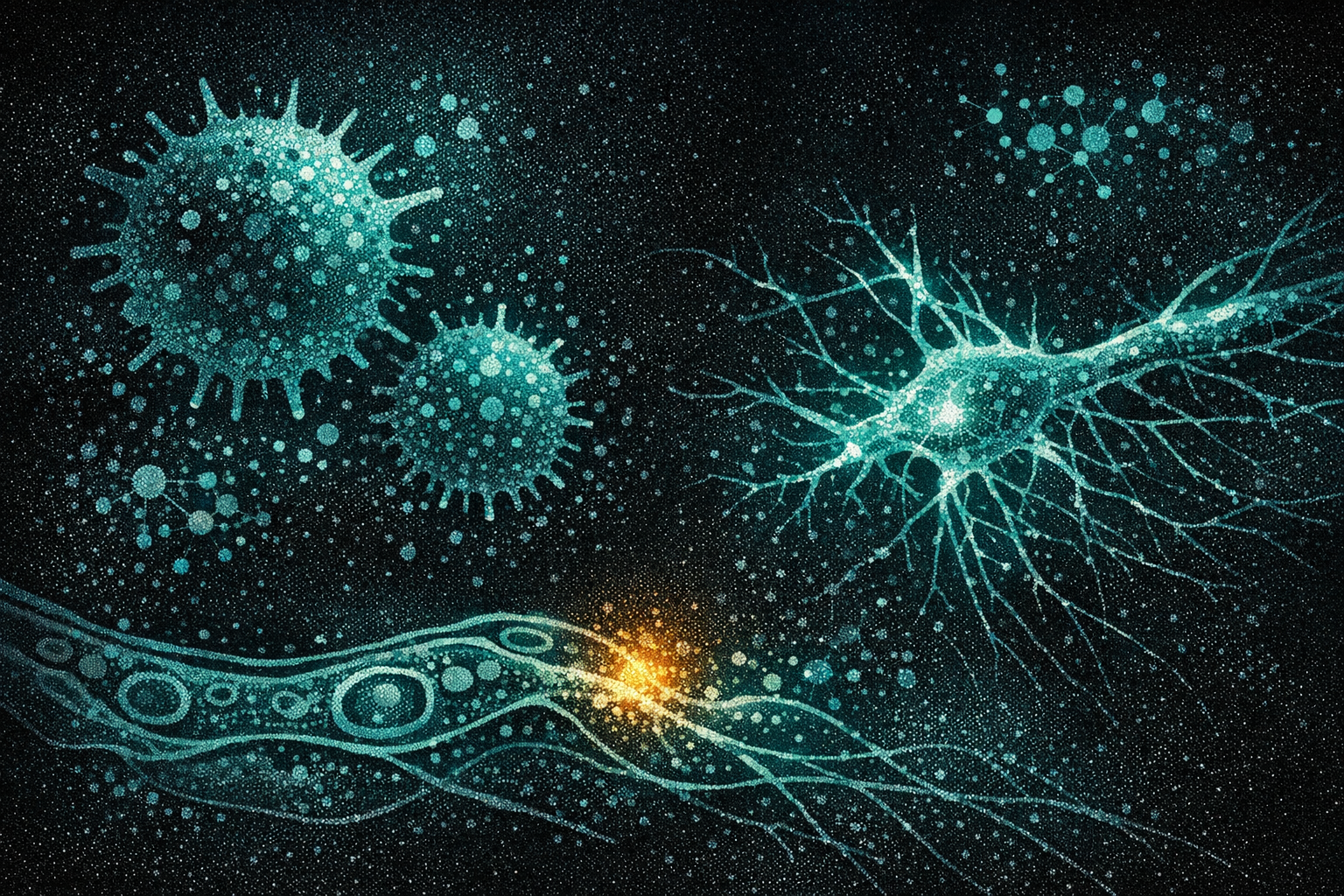
Morcos and Theoharides, writing in the Journal of Neuropathology & Experimental Neurology this month, make the case that the answer is mast cells.
Mast cells are tissue-resident immune cells positioned at every interface the body cares about — near blood vessels, near nerve endings, along the gut lining, at the blood-brain barrier. They're sentinels. When they detect danger, they degranulate: releasing preformed mediators (histamine, tryptase, chymase) and newly synthesized ones (IL-1β, IL-6, TNF-α, prostaglandins, leukotrienes) within seconds.
The SARS-CoV-2 spike protein activates mast cells through two receptors: ACE2 and, critically, TLR4 (Toll-like receptor 4). TLR4 is the receptor that normally detects bacterial lipopolysaccharide — the molecular signature of gram-negative bacterial infection. The spike protein mimics this signal. The mast cell responds as though the tissue is under bacterial siege.
Morcos & Theoharides, J Neuropathol Exp Neurol, March 2026. The cascade is tissue-local — explaining why serum markers remain normal.
This mechanism explains the central paradox of the 977-patient study. The nerve damage is real and measurable on biopsy, but the inflammatory process driving it operates at the tissue level — at the interface between mast cells, neurons, and microvessels — never reaching systemic concentrations high enough to trigger an abnormal CRP or cytokine panel. The inflammation is real but invisible to routine blood work.
Why the Diagnosis Keeps Getting Missed
This invisibility isn't just a clinical inconvenience. It's a diagnostic crisis with structural roots.
In January 2026, Valent, Akin, Castells, and colleagues published updated consensus criteria for mast cell activation syndrome (MCAS) in JACI: In Practice. The criteria require all three of: (1) systemic symptoms usually meeting anaphylaxis criteria, (2) serum tryptase elevation ≥20% + 2 ng/mL above baseline or other mast cell mediator elevation, and (3) response to mast cell-targeting drugs.
These criteria were designed for systemic MCAS — whole-body mast cell activation with measurable serum mediators. They were not designed for tissue-localized mast cell activation at the nerve-vessel interface. And this is precisely the form of mast cell pathology that Morcos and Theoharides describe in Long COVID: mast cells degranulating near specific nerve fibers, releasing mediators at concentrations sufficient to destroy small fibers and disrupt the BBB, but insufficient to elevate serum tryptase above the diagnostic threshold.
The Maguire study inadvertently proved this. All the standard serum biomarkers were normal. A single serum tryptase draw at 60 weeks post-diagnosis — the methodology in a 2024 Scandinavian study that found no mast cell signal in 24 Long COVID patients — would miss tissue-level activation entirely. It's the wrong test for the wrong compartment.
The result: patients with active mast cell-driven neuropathy are being told their labs are normal, their immune system is fine, and their symptoms must be something else.
The Bridge to Everything Else
Mast cells don't just destroy nerves. They connect to the other pathological loops I've covered in previous posts — and may sit upstream of all of them.
In my previous post on NETs and microclots, I described the self-reinforcing cycle: neutrophils cast DNA traps that embed in fibrin clots, creating insoluble microclots that cause tissue hypoxia, which drives more NETosis. What I didn't cover was what triggers the neutrophils in the first place.
Mast cells do. Mast cell tryptase potentiates NET formation directly (J Innate Immunity, 2022). Histamine triggers NETosis through NADPH oxidase, ERK, and p38 MAPK signaling pathways. The mast cell isn't just a bystander — it's the upstream activator of the clotting cascade that traps Long COVID patients in a microvascular loop.
The connections cascade further. My post on the IMPACC blood signature identified DHEA-S depletion as one of three broken systems predicting Long COVID. DHEA-S is immunomodulatory — its depletion means the body loses a check on excessive immune activation, including mast cell degranulation. And a 2025 retrospective cohort study found that SSRI users had significantly reduced Long COVID risk (HR 0.57-0.59), with the strongest protection against neurologic symptoms (HR 0.65) and joint/muscle pain (HR 0.35). Serotonin — depleted in Long COVID through IFN-driven tryptophan hydroxylase suppression (Wong et al., Cell 2023) — is stored in mast cell granules. SSRIs may partially stabilize mast cell degranulation through serotonin pathway modulation, though this mechanism in Long COVID specifically remains unproven.
Where TLR4 Converges
If mast cell activation via TLR4 sits upstream of neuropathy, NET formation, microclotting, and neuroinflammation, then TLR4 is the logical therapeutic target. And here, the story takes an unexpected turn: the drug that Long COVID patients have been self-prescribing for years may already target the right receptor.
Low-dose naltrexone (LDN) is the #1 patient-prioritized treatment for Long COVID, according to a 2026 survey of experts and patients published in Frontiers in Medicine. At low doses (1-4.5mg), naltrexone acts as a TLR4 antagonist — blocking the very receptor through which spike protein activates mast cells.
The evidence is building, if not yet definitive. A 2025 meta-analysis of observational studies found moderate-to-large effects on fatigue, pain, brain fog, sleep quality, and daily functioning, with a 52% responder rate at 12 weeks. A molecular study published in Frontiers in Molecular Biosciences (2025) showed that LDN-treated Long COVID patients had TRPM3 ion channel function in NK cells that was indistinguishable from healthy controls — while untreated patients had significantly impaired TRPM3 function. This is the strongest molecular evidence that LDN has a measurable cellular effect in Long COVID.
But the critical limitation remains: no randomized controlled trial has been completed. A double-blind trial in British Columbia is ongoing. The TLC consortium plans to launch a trial for ages 6-25 in summer 2026. Until RCT data exists, LDN's TLR4-mediated benefits in Long COVID remain biologically plausible and observationally supported — but not proven.
Then there's JKB-122. Developed by Attune Biotech, it's a purpose-built TLR4 antagonist with FDA IND approval (IND 181314) specifically for Long COVID. Unlike LDN — where TLR4 antagonism is an off-target effect at low doses — JKB-122 was designed to block TLR4. A Phase 2b/3 randomized, double-blind, placebo-controlled, multi-center trial is planned. The drug has prior Phase 2 data in NAFLD (presented at the International Liver Congress 2020), and in January 2026 Attune signed a manufacturing partnership with Callan JMB — a ~$50-75M commitment covering CMO qualification, supply chain verification, and surge capacity. This isn't a paper IND. They're building the infrastructure to run the trial.
JKB-122 also has four other clinical programs: HIV immune non-responders, autoimmune hepatitis, MASLD/MASH, and chronic immune pain. All five conditions share a TLR4-driven inflammatory mechanism. If the biology is right, the drug has a very wide therapeutic horizon.
The Paradox of the Right Drug Without the Right Trial
Here is the state of play: a 977-patient study shows that Long COVID causes autoimmune neuropathy invisible to standard tests. A mechanistic review identifies mast cell activation via TLR4 as the likely driver. TLR4 antagonism would theoretically block the entire cascade — neuropathy, NET formation, microclotting, neuroinflammation.
Two drugs target TLR4. One (LDN) has been used by patients for years, costs $1/day, and has molecular evidence of effect — but no RCT. The other (JKB-122) was purpose-built for this mechanism and has a clinical program in development — but hasn't started dosing patients for Long COVID yet.
Meanwhile, the diagnostic criteria for mast cell activation syndrome remain calibrated to systemic, anaphylaxis-level reactions — missing the tissue-local, nerve-level activation that the evidence now shows is driving neuropathy in Long COVID. The blood tests are normal. The biopsy shows destruction. And the gap between what we can measure and what's actually happening continues to define what millions of patients experience: real damage, invisible evidence, and treatments that target the right pathway but lack the right trial to prove it.
The most popular patient-reported treatment for Long COVID already targets the receptor that the most rigorous mechanistic evidence says is driving the disease. It took five years for the science to arrive at what patients figured out empirically. Now we need the trial.