Your body is full of microscopic traps. Not in your arteries — a cardiologist could see those. These are in your capillaries, the smallest blood vessels in your body, each narrower than a human hair. They're made of DNA, enzymes, and proteins that were supposed to protect you. Instead, they're slowly strangling your tissues.
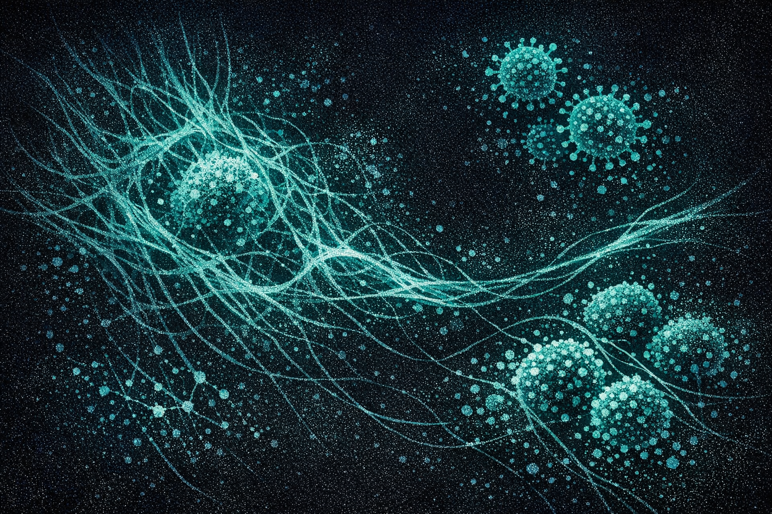
In healthy people, neutrophils — the most abundant white blood cells — cast webs of chromatin and antimicrobial proteins into the bloodstream to catch and kill pathogens. These neutrophil extracellular traps, or NETs, are an ancient defense mechanism. They're elegant, efficient, and self-limiting. The body deploys them, they trap bacteria, and then enzymes dissolve them. The system resets.
In Long COVID, the system doesn't reset. The NETs don't dissolve. They embed themselves in fibrin clots, stabilizing structures that were supposed to be temporary into something permanent. And for the first time, researchers have shown exactly how this happens — and why it creates a feedback loop the body cannot escape.
The Discovery
In November 2025, a collaboration between Thierry Bagot's team in Montpellier and Etheresia Pretorius's group in Stellenbosch published what may be the most important clotting paper in Long COVID to date. They recruited 50 Long COVID patients and 38 healthy controls, then examined their blood with fluorescence microscopy and machine learning.
NETs and Microclots in Long COVID
The numbers alone are striking: microclots were nearly 20 times more abundant in Long COVID blood. But the real breakthrough was what the microscopy showed. Using fluorescent markers for both fibrin (the structural protein of clots) and NET components (DNA, myeloperoxidase, neutrophil elastase), the team demonstrated something never before reported: NETs were physically embedded within the microclots. Not adjacent to them. Not floating near them. Woven into their fibrin architecture.
This structural integration is critical. Normal fibrin clots are dissolved by plasmin, an enzyme the body produces specifically for this purpose. But when NETs infiltrate the clot structure, their histones and DNA physically shield the fibrin from plasmin access. As Pretorius stated: the NET-microclot interaction "could render microclots more resistant to fibrinolysis, promoting their persistence in circulation."
The body is building clots it cannot dissolve.
How COVID Starts the Chain
The pathway from SARS-CoV-2 infection to persistent NETosis involves a surprisingly clean mechanistic chain — one that doesn't require the virus itself to persist in the body.
It starts with platelets. The SARS-CoV-2 spike protein activates platelets through CLEC-2, a C-type lectin receptor, via Syk kinase signaling. Activated platelets don't just clump — they release tiny packages called extracellular vesicles (EVs) loaded with microRNAs, particularly miR-21 and let-7b.
These platelet EVs are the messengers. When they reach neutrophils, the microRNAs activate TLR7/8 (toll-like receptors that normally detect viral RNA). This triggers phosphorylation of p47phox, assembly of the NADPH oxidase complex, and a burst of reactive oxygen species. The ROS overwhelm the neutrophil, causing it to expel its chromatin — a controlled cell death that creates the NET.
Each step is well-documented in separate papers. What hasn't been appreciated until now is how cleanly they connect: spike activates platelets, platelets program neutrophils, neutrophils cast NETs, NETs stabilize clots. And the spike protein doesn't need to be freshly produced by an active infection. Post-acute coagulopathy research has shown that spike protein expression can persist in endothelial cells and platelets long after the acute phase, and even antibody-bound spike fragments retain the ability to activate CLEC-2.
The Loop That Won't Break
If the chain stopped at NET-stabilized microclots, the problem would be serious but potentially manageable. What makes it devastating is what happens next.
Microclots lodge in capillaries. They reduce blood flow. Tissues downstream become hypoxic — starved of oxygen. And in 2025, Ye and colleagues published the missing piece of the puzzle in Frontiers in Immunology: hypoxia itself drives more NETosis.
The mechanism is specific and now fully mapped. When oxygen levels drop, cells stabilize a transcription factor called HIF-1α (hypoxia-inducible factor 1-alpha). In neutrophils, HIF-1α reprograms energy metabolism from oxidative phosphorylation to glycolysis. This metabolic switch — enhanced glycolysis under hypoxic conditions — directly promotes NET formation. Ye's team showed that both LW6 (a HIF-1α inhibitor) and Bay-876 (a glycolysis blocker) reduced NETosis in vitro, confirming the causal chain.
This is the core of the problem. NETs create microclots. Microclots cause hypoxia. Hypoxia, through HIF-1α-dependent glycolytic reprogramming, creates more NETs. Each revolution of the cycle adds more insoluble clots to the circulation and deepens the oxygen debt in affected tissues. The loop is self-amplifying, and the body has no built-in mechanism to break it once it starts.
There's an additional amplifier: the spike protein increases P-selectin expression on endothelial cells, which recruits more neutrophils to vessel walls — providing fresh fuel for the NETosis engine. And as I covered in my previous post on co-infections, Candida biofilms in the hypoxic gut create their own microenvironments that push neutrophils toward NETosis, adding yet another feed into the loop.
What This Feels Like
The clinical consequences map directly to what Long COVID patients experience. Research from the Wüst group at VU Amsterdam, published in Medicina (2024), has documented reduced capillary density and impaired mitochondrial function in ME/CFS patients — a condition that shares extensive pathophysiology with Long COVID.
In their model, all degrees of capillary obstruction can coexist simultaneously: some capillaries merely slowed, others in stasis, others fully blocked. The result is a patchwork of tissue perfusion — some areas getting adequate oxygen, others chronically starved. This explains several hallmark Long COVID symptoms:
Symptoms Explained by Microvascular Obstruction
This is why "just push through it" is dangerous advice. When patients with post-exertional malaise exercise, they're forcing oxygen-starved tissue to work harder — deepening the hypoxic drive that feeds NETosis. Exercise doesn't break the loop. It accelerates it.
Toward a Blood Test
One of the most immediately practical findings from the Thierry-Pretorius paper is the diagnostic potential. Their machine learning model, trained on microclot and NET markers, achieved 95% diagnostic accuracy for identifying Long COVID patients from blood samples — 91% in blinded validation. The combination of MPO (myeloperoxidase) and small microclot counts reached 98% specificity.
This matters enormously. Long COVID currently has no objective diagnostic test. Patients are diagnosed by symptom checklists and exclusion of other conditions — a process that's slow, subjective, and frequently dismissive. A blood-based biomarker panel measuring NET components and microclot density could provide the objective confirmation that millions of patients need, and that clinicians, insurers, and disability systems demand.
The test isn't available yet. It requires fluorescence microscopy and computational analysis that most clinical labs don't have. But the path from research tool to diagnostic assay is far shorter than the path from drug target to approved treatment.
The Treatment Gap
Here is the honest state of therapeutics targeting this pathway. It is not encouraging.
| Approach | Target | Evidence | Status |
|---|---|---|---|
| Nattokinase | Fibrin microclots | 70-80% reduction in vitro | No clinical trial |
| Lumbrokinase | Fibrin microclots | Pilot trial completed | Results pending |
| Triple anticoagulation | Clotting cascade | 91 pts, uncontrolled | No RCT, not peer-reviewed |
| DNase I (dornase alfa) | NET DNA | Two RCTs failed (acute COVID) | Delivery problem |
| PAD4 inhibitors | NETosis (upstream) | JBI-589 preclinical | No human trials |
| HIF-1α inhibitors | Hypoxic NETosis | LW6 in vitro | Speculative |
Nattokinase showed the most impressive preclinical results. A collaboration between the University of Liverpool and Stellenbosch (Kell & Pretorius, 2024) used automated fluorescence microscopy to show that nattokinase dismantled the amyloid fibrin core of microclots — reducing fluorescent intensity by 70-80% within two hours in vitro. But no randomized clinical trial has been conducted in Long COVID patients.
The lumbrokinase pilot trial (NCT06511050), funded by PolyBio Research Foundation at Mount Sinai, completed its expected enrollment period in February 2026. It tested six weeks of lumbrokinase in patients with Long COVID, ME/CFS, and long Lyme disease, measuring microclots before and after treatment. Results have not yet been published — they may be the first clinical evidence for fibrinolytic therapy in post-viral microclotting.
DNase I (dornase alfa), which degrades NET DNA, failed in two acute COVID trials — a Swedish study of 76 patients and a French trial of 77 ARDS patients. The problem appears to be delivery: aerosolized DNase reaches the lungs but not the systemic circulation where NET-stabilized microclots reside. Intravenous DNase has not been tested. No LC-specific trial exists.
PAD4 inhibitors target the upstream machinery of NETosis itself. PAD4 (peptidylarginine deiminase 4) citrullinates histones, causing chromatin decondensation — the critical step before a neutrophil expels its DNA as a NET. JBI-589 from Jubilant Therapeutics is the most advanced: oral, selective, and potent in preclinical arthritis models. Ten companies hold 28 patent applications in this space. But no PAD4 inhibitor has entered human clinical trials for any indication as of March 2026.
The most controversial approach is Pretorius and Laubscher's triple anticoagulation protocol — dual antiplatelet therapy (clopidogrel + aspirin) plus a direct oral anticoagulant (apixaban) with a proton pump inhibitor. In an uncontrolled case series of 91 Long COVID patients, they reported symptom resolution in the majority and microclot score drops from 7.1 to 5.2, with no serious bleeding events. But without randomization, blinding, or a control group, these results cannot be interpreted as causal evidence. No RCT has been conducted.
Breaking the Loop
The central insight of the NETs-microclots research is that this is not a linear problem with a single point of failure. It's a cycle with at least four distinct nodes: platelet activation, NETosis, microclot stabilization, and hypoxic amplification. Addressing only one node may slow the loop but cannot stop it.
Dissolving clots (nattokinase, lumbrokinase) doesn't prevent new NETs from forming. Blocking NETosis (PAD4 inhibitors) doesn't dissolve existing clots. Preventing platelet activation doesn't reverse established hypoxia. And improving oxygenation doesn't clear the structural traps already embedded in fibrin.
The loop must be broken at multiple points simultaneously. This is the fundamental challenge — and the reason that five years into the Long COVID crisis, not a single approved therapy targets the clotting pathology that may underlie its most disabling symptoms.
A 95% diagnostic test exists in a research lab. A 70-80% clot-dissolving enzyme works in a dish. A pathway-specific NETosis blocker shows preclinical efficacy. The biological understanding is arguably ahead of any other Long COVID mechanism. What's missing is the clinical infrastructure to turn these findings into treatments — and the funding to run the combination trials this biology demands.
The blood won't flow on its own. Something has to break the loop.